SERVICES
Canine Forelimb/Elbow Injuries
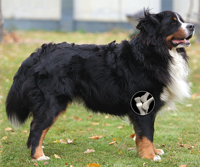
The elbow joint is a very complex structure. Each of the three bones which make up the joint contribute significantly to the integrity of a highly moveable, complicated structure. There are a series of common problems that are seen in the elbow joint of large breed, young dogs. If they are not detected or corrected at a young age, arthritis often develops. Correcting the problem early will address the lameness that is present as well as limit the amount of arthritis that will develop.
The long term effect of these conditions and abnormal development is degenerative joint disease (arthritis) leading to wear and erosion of the cartilage on the medial aspect of the elbow joint. Over time, the cartilage deterioration causes increasing inflammation, joint instability and severe pain. The resultant syndrome is referred to as Medial Compartment Disease. Medial Compartment Disease has long been difficult to effectively treat. Although pain management drugs may help the dog feel better and cope with a bad elbow, they do not alter the progression of the disease.
We have a great deal of experience with orthopedic conditions of the elbow. Treatment options depend upon the diagnosis and prognosis of the individual patient but may include the following:
Click on the procedure below to learn more.
Osteochondritis Dissecans (OCD)
OCD is a cartilage disorder that can affect any joint. It is most common in large breed, fast growing dogs like Labrador and Golden Retrievers. OCD occurs when a separation develops between the cartilage surface within the joint and the abnormal subarticular cartilage and underlying bone. When the separation occurs, a blister-like lesion develops on the cartilage surface. Often, a crack will form around the edge of the lesion and allow joint fluid to leak into the lesion and create a flap. This causes pain to the patient (like having a constant "pebble in your shoe")and sets up inflammation within the joint which leads to arthritis.
The typical age of initial onset of OCD lameness symptoms is 4-10 months. Early diagnosis and intervention is the key to preventing long-term irreversible damage to the joint. Correction of the problem requires surgical removal of the OCD lesion. Once the lesion has been removed, the body will fill the area in with a different type, but similarly functional, form of cartilage. Patients with chronic OCD will see improvement of symptoms with surgery, but the presence of significant arthritis in the joint may still cause occasional lameness.
Ununited Anconeal Process (UAP)
The Anconeal Process is a part of the ulna that, when the elbow joint is extended, helps to stabilize the joint. It has it's own growth plate from which the bone grows, which typically closes by about 20 weeks of age. If the growth plate remains open beyond 20 weeks, it is considered "ununited". The significance of this is that the Anconeal Process is detached from the main piece of bone and is essentially a loose piece that is unstable within the joint. Surgery should be performed to address and remove the unattached piece but, even with surgical intervention, the elbow joint is still not "normal" and is considered compromised to some degree. Surgery slows the progression of arthritis, but will not prevent it entirely.
Fragmented Coronoid Process (FCP)
The Coronoid Process is another prominence of bone on the ulna in the elbow joint. It also has its own area of bone growth, similar to the Anconeal Process. The Coronoid Process is often difficult to visualize on radiograph, and often requires specialized positioning and radiograph exposure techniques. Similar to UAP, an FCP creates a loose piece of bone within the joint. Given the complex nature of the elbow, having a loose piece of bone in the joint initiates significant arthritic changes very quickly. FCP is a condition that warrants prompt, aggressive surgical intervention.
Proximal Abducting Ulna Osteotomy (PAUL)
Studies suggest that dogs bear a majority of their weight on the medial (inner) aspect of the elbow joint. In dogs with Medial Compartment Disease, this unequal load on the deteriorating cartilage of the elbow joint causes debilitating pain. With a long history of ineffective treatment options, the PAUL procedure is a novel and exciting new surgical technique for the treatment of dogs with Medial Compartment Disease. In essence, this procedure allows us to make a subtle change in limb alignment which unloads the medial compartment and makes better use of the healthy lateral (outer) cartilage surface. The PAUL procedure has shown to be a viable treatment option to help treat and prevent the devastating effects of this disease.
CUE (Canine Unicompartmental Elbow) Procedure
Another way of approaching advanced medial compartment disease of the elbow is the CUE procedure. This advanced surgical approach involves replacing the damaged joint surfaces of the inner (medial) portion of the elbow joint with implants in a partial joint replacement procedure. The implants provide new artificial joint surfaces where the cartilage has been worn away, relieving pain and improving function of the limb.
Fracture Repair
At Packerland Veterinary Center, we are able to handle the simplest of broken bones, all the way through the most complex of fracture presentations. Dr. Dunbar's extensive training and orthopedic experience have given him the tools to provide the best care possible for fractures of all shapes and sizes.
There is no cookie cutter method to approach every patient with a broken bone. We have to be flexible in the methods that we have available to approach each individual situation. Some fractures may heal adequately with external support such as casting, while others require elaborate bone plate and screw repairs along with bone grafting techniques. The trick is to know what works with each fracture presentation, and to be able to perform the procedure in a manner that will give the patient the best recovery possible.
Stem Cell Therapy (SCT)
This procedure involves surgically harvesting fat tissue from your pet and then processing that tissue in house to remove the stem cells. The stem cells are then "activated" with our special equipment and later the same day we can inject the cells back into specific areas of the patient where needed. Stem Cells will not change any structural issues that have lead to any current elbow problems, but can help with the arthritis that has formed in the joint by helping to reduce inflammation and rebuild the joint cartilage surface, making the pet more comfortable overall. In many cases, SCT can often be used in conjunction with surgical treatment to maximize overall results.
For more in-depth information about Stem Cell Therapy, click here.
Platelet Rich Plasma Treatment (PRP)
Popular in human orthopedics, PRP therapy utilizes a patient's own blood plasma as an all-natural, drug free pain relief option. Once activated by our specialized equipment, the PRP works to initiate and accelerate tissue repair and reduce localized inflammation. PRP can be utilized in a variety of conditions, but most often is used to treat the inflammation present in injured joints and tendons. While it does not provide a more long-term regeneration of cells like Stem Cell Therapy, PRP can be an affordable short term solution to treat pain and inflammation in our patients.
PRP treatment begins with a blood draw from the patient. The blood is then specially processed to bring out the PRP mix of concentrated serum and platelets. Patients then receive light sedation while the PRP is injected into the specific area or areas needing treatment. Any additional PRP can be frozen and saved short-term for future treatments that may be needed in the months following. While each pet's response to PRP is different, we have seen great short and longer term results utilizing this therapy.