
SERVICES
Canine Shoulder Injuries
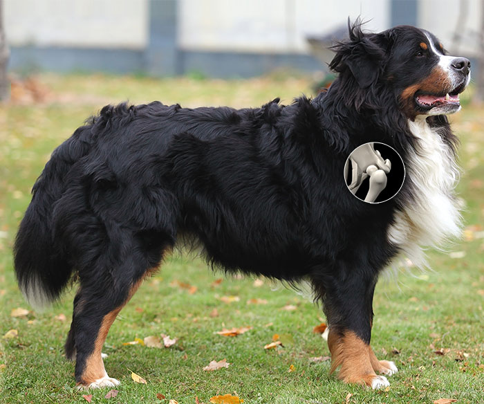
Lameness of the forelimbs can have many causes, some of which are not immediately obvious and require more in-depth investigation. The shoulder is a very complex joint in which we can see a variety of conditions. Many conditions can be diagnosed using thorough examination and radiographs, but others may require different methods. If we suspect a particular joint is causing discomfort, but are not able to confirm via objective methods such as X-ray, we may utilize intra-articular injection as a diagnostic tool. In these cases we inject a steroid into a suspected problematic joint. The pet's response to this medication gives us a great deal of information on how to proceed with our treatment. Additionally, we have the ability to utilize our ultrasound unit to help us take a closer look at the tendons of a joint without performing surgery. When the shoulder can be pinpointed as the problem area, there are a few common conditions we treat.
Click on the procedure below to learn more.
Osteochondritis Dissecans (OCD)
OCD is a cartilage disorder that can affect any joint and is the most common orthopedic condition we see within the shoulder joint in young dogs. It is most common in large breed, fast growing dogs like Labrador and Golden Retrievers. OCD occurs when a separation develops between the cartilage surface within the joint and the abnormal subarticular cartilage and underlying bone. When the separation occurs, a blister-like lesion develops on the cartilage surface. Often, a crack will form around the edge of the lesion and allow joint fluid to leak into the lesion and create a flap. This causes pain to the patient (like having a constant "pebble in your shoe") and sets up inflammation within the joint which leads to arthritis.
The typical age of initial onset of OCD lameness symptoms is 4-10 months. Early diagnosis and intervention is the key to preventing long-term irreversible damage to the joint. Correction of the problem requires surgical removal of the OCD lesion. Once the lesion has been removed, the body will fill the area in with a different type, but similarly functional, form of cartilage. Patients with chronic OCD will see improvement of symptoms with surgery, but the presence of significant arthritis in the joint may still cause occasional lameness.
Biceps Tendon
Bicipital Tenosynovitis, or inflammation of a tendon and its sheath, is a very painful condition most often seen in older medium and large size dogs. The exact cause of the condition is unknown, but several possibilities include: tendon injury, involvement secondary to arthritis or other orthopedic conditions such as OCD, and infection. Classic presentation includes intermittent forelimb lameness which worsens after activity. Examination of these patients typically reveals discomfort with deep palpation over the area of the biceps tendon.
There are a variety of ways to approach a case of Bicipital Tenosynovitis. Some patients respond fairly well to anti-inflammatory medications, intra-articular steroid injections or even PRP treatments. If these become less effective or the patient's symptoms are more severe in nature, surgery may be needed to relieve patient discomfort. Surgery can often be done arthroscopically which minimizes tissue injury and results in a faster return to function. There are a variety of techniques that can be utilized in surgery, and the approach is customized for each patient which optimizes their prognosis and recovery.
Arthroscopy
Arthroscopy is a minimally invasive procedure that allows our veterinarians to view the internal environment and structures of joints and in some cases perform corrective procedures. This technology utilizes a small scope attached to a camera and viewing screen that is inserted within the joint to allow the surgeon the view the joint environment.
Arthroscopy is beneficial because it allows direct visualization of the cartilage surfaces and soft tissue under magnification. Due to the relatively small size of the instruments, there is often less tissue injury. Minimal tissue disruption causes less post-operative discomfort for the patient, and typically less scar tissue development. Patients undergoing arthroscopic procedures typically have a shorter recovery time and quicker return to function. When appropriate, patients requiring shoulder surgery can often benefit from the use of Arthroscopy.
Stem Cell Therapy (SCT)
This procedure involves surgically harvesting fat tissue from your pet and then processing that tissue in house to remove the stem cells. The stem cells are then "activated" with our special equipment and later the same day we can inject the cells back into specific areas of the patient where needed. Stem Cells will not change any structural issues that have lead to any current shoulder problems, but can help with the arthritis that has formed in the joint by helping to reduce inflammation and rebuild the joint cartilage surface, making the pet more comfortable overall. In the case of shoulder issues, SCT is often used in conjunction with surgical treatment to maximize overall results.
For more in-depth information about Stem Cell Therapy, click here.
Platelet Rich Plasma Treatment (PRP)
Popular in human orthopedics, PRP therapy utilizes a patient's own blood plasma as an all-natural, drug free pain relief option. Once activated by our specialized equipment, the PRP works to initiate and accelerate tissue repair and reduce localized inflammation. PRP can be utilized in a variety of conditions, but most often is used to treat the inflammation present in injured joints and tendons. While it does not provide a more long-term regeneration of cells like Stem Cell Therapy, PRP can be an affordable short term solution to treat pain and inflammation in our patients.
PRP treatment begins with a blood draw from the patient. The blood is then specially processed to bring out the PRP mix of concentrated serum and platelets. Patients then receive light sedation while the PRP is injected into the specific area or areas needing treatment. Any additional PRP can be frozen and saved short-term for future treatments that may be needed in the months following. While each pet's response to PRP is different, we have seen great short and longer term results utilizing this therapy.